All Medical Services

Featured by Department

Urologist / Uro-Surgeon

A urologist is a physician who has specialized knowledge and skill regarding problems of the male and female urinary tract and the male reproductive organs. He deals with diseases of whole genitourinary system, start from kidney, bladder, prostate, testis, penis to female genitalia. Any problem of urinary system or genital system are best taken care of by a urologist.

Few examples are :

  • Prostate Enlargement (BPH)
  • Kidney, Ureter and Urinary bladder stone
  • Recurrent urinary tract infection both male and female
  • Blood in urine or semen
  • Increased urinary frequency both day and night
  • Overactive Bladder
  • Thin stream of urine
  • Bedwetting by children
  • Male sexual and infertility problems
  • Cancer of Kidney, Adrenal, Urinary Bladder, Prostate, Testis and Penis
  • Congenital (since birth) urinary disease- Paediatric urology
  • Uro-Gynaecology like urine leakage in female

+91- 9798900124

Dr Sanjay Jouhary

(MBBS, MS, DNB, Urology)

post_502443_1485782625

What is the prostate gland?

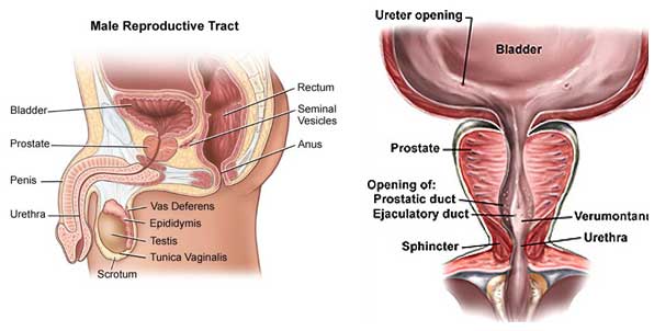
The prostate is a walnut (अखरोट) sized gland weigh around 15-20 gram. It is part of male reproductive system. It is located below the urinary bladder and surrounds the upper portion of the urethra (the tube that carries urine out of the body). Its growth is dependent on male hormone Testosterone

What is its function?

The prostate gland makes the fluid portion of the semen. This fluid energizes the sperm and makes the vaginal canal less acidic.

What is benign prostatic hyperplasia (BPH) ?

The prostate gland begins to enlarge after 40 years of age. About half of all men between the ages of 50 and 60 have BPH. Up to 90% of men over age 80 have BPH. The reason for the enlargement is not fully clear, and is likely to be due to some hormonal changes on aging. This enlargement is not due to cancer.

What are the consequences of this enlargement?

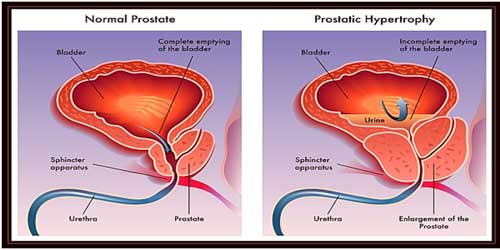
The prostate tissue as it enlarges squeezes the urethra, creating a mechanical obstruction to emptying of the bladder. Initially the bladder respond by contracting more strongly so that the urine can be emptied and the patient has no symptoms except slight diminution of flow. Later the obstruction causes irritability of the bladder muscle causing increased urinary frequency day & night and urgency. As the disease progresses, the bladder weakens, resulting in incomplete bladder emptying, eventually progressing to stoppage of urinary flow and  retention of urine. Sometimes the high pressures generated in the bladder due to obstruction are transmitted back to the kidneys. This is a potentially dangerous situation, and can result in serious kidney damage.

post_48895_1485777504 (1)

What are the symptoms of BPH?

The most common symptoms of BPH are :

Increased urinary frequency both day and night.

  • Urgency -Feeling that urinating can’t wait
  • Weak urine flow
  • Dribbling of urine
  • The need to stop and start urinating several times
  • Trouble starting to urinate
  • The need to push or strain to urinate
  • Sense of incomplete empty

Does the size of the prostate determine severity of symptoms?

The size of the prostate gland does not always correlate with severity of obstruction and symptoms . Some men with greatly enlarged glands have little obstruction and few symptoms while others, whose glands are less enlarged, have more blockage and greater problems.

What causes acute urinary retention?

In a patient of BPH with partial obstruction, severe constipation, alcohol, cold temperatures, or a long period of immobility can bring on urinary retention. Certain drugs like cough syrups and cold remedies may also precipitate urinary retention.

post_508652_1485782666

How is BPH diagnosed?

  • AUA/IPSS Symptom score
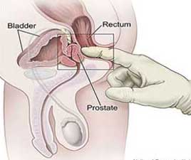
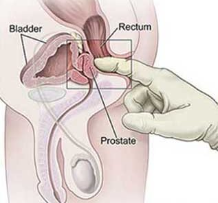
  • Digital rectal examination – The doctor inserts a gloved finger into the rectum and examine the prostate gland next to the rectum, and get a general idea of its size and condition.
  • Urinanalysis
  • Serum PSA
  • Uroflowmetry
  • Ultrasonogrphy of kidney, ureter, bladder, prostate and post void tesidual urine volume

What are the complications of BPH?

  • Recurrent urinary tract infection.
  • Urinary retention
  • Bladder and kidney damage.
  • Bladder stone.
  • Haematuria- blood in urine

If the bladder is permanently damaged, treatment for BPH may be ineffective. When BPH is found in its earlier stages, there is a lower risk of developing such complications. So sooner the treatment started better is the outcome

Does BPH cause cancer?

BPH does not cause cancer. However, cancer does occur more often as men get older. Therefore, it is important that the prostate gland is checked regularly. All men over age 50 should have an annual prostate examination and those who have a higher risk because of a family history should be examined yearly starting at age 40.

what are the treatment?

1. life style modification

2. Medical treatment

3. Surgical treatment

What are the oral medications available?

The prostate gland is made up of muscles and glands, There are mainly two groups of drugs available that work on these tissues.

1. Alpha-blocking drugs such as Tamsulosin, relax the muscles at bladder neck and prostate and helps in improving voiding symptoms. These drugs have certain side effects like dizziness, tiredness, as they tend to lower blood pressure also retrograde ejaculation of semen.

2. 5-Alpha reductase inhibitors like finasteride inhibit production of the hormone DHT, which is involved with prostate enlargement. Up to 20-30 % reduction in prostate size may be achieved with this agent in 6 months. Patients get symptomatic relief and risk of complications like acute urinary retention is reduced. It is important to realise that these drugs do not cure BPH: they only alleviate symptoms, so to be taken for life.

3) Anti-muscarinic agents- these drugs act on detrussor muscle of bladder and relieves storage symptoms

What are the surgical options?

In older days the surgical process involved “cutting & stiching” procedure (open surgery) with associated blood loss, higher complications, prolonged hospitalisation. Nowadays the technique of transurethral surgery (popularly known as “microsurgery”) has made the surgery a lot simpler. This is a safe and effective operation performed in large numbers of patients with good results.

Transurethral surgery ( Transurethral resection of prostate : TURP )

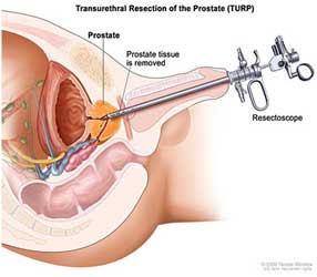
This type of surgery is popularly known as microsurgery, it is one of the  common surgery done world wide for prostate enlargement. In this surgery there is no external cutting/incision or stitching. This operation is usually performed in spinal anaesthesia by giving one injection on back, that means patient is not fully anaesthetised, only lower half of his body is made senseless, patient can watch his surgery and even can talk with his doctor. Doctor introduces a fine instrument called resectoscope through patients urethral, reaches up to prostate and a wide channel for urinary passage is made under vision at the level of obstructing prostate. The resected prostatic tissues are removed through same opening. Hospital stay after this surgery is usually 2-3 days.

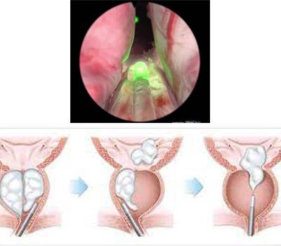
Till recently TURP was considered world wide “GOLD STANDARD” procedure for prostate surgery because of its high success rate, minimal complication rate, quick procedure, short hospital stay and cost effectiveness. But since the advent of laser surgery ThuFLEP ( Thulium Fiber Laser Enucleation Of Prostate) is now considered world wide new Gold Standard Surgery for enlarged Prostate.

TURP can be performed in any age group patient after anaesthesia fitness, it can be performed in patients with hypertension, diabetes mellitus or kidney disease.

post_984228_1485778512

ThuFLEP – Thulium Fiber Laser Enucleation Of Prostate

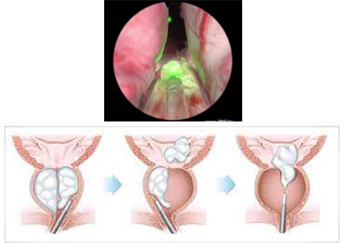
 ThuFLEP ( Thulium Fiber Laser Enucleation Of Prostate) is considered world wide new Gold Standard Surgery for enlarged Prostate because of highest success rate. This is most advanced and recent technique for management of enlarged prostate. It has minimum or almost no complications, There is no risk of electrolyte imbalance in it ( which is possible in TURP)

ThuFLEP can be performed in any old age group person, even heart disease persons with pacemaker are not contraindicated. There is negligible or no bleeding and other complications with ThuFLEP. Hospital stay is only 1-2 days. Catheter duration is usually 48 hours.

Laser1

What lifestyle modifications are advised in a patient with BPH?

When symptoms of BPH are not severe, these lifestyle changes might be helpful.
  • Limit intake of tea, coffee, and alcohol. Alcohol increases urine production and may cause congestion in the prostate gland. Caffeine irritates the bladder.
  • Stay active, Urine is retained when you don’t move around.
  • Stay warm, cold can precipitate retention of urine.
  • Avoid over-the-counter drugs of cold and cough. These drugs tighten the muscles that control urine flow, making it more difficult to urinate.
  • Restrict fluid intake after 6 p.m.
  • Each time you urinate, try to empty your bladder completely.
  • Avoid costipation
post_160264_1486199285

Kidney Stone

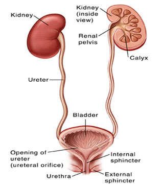
The urinary tract consists of a pair of kidneys which are two bean shaped organs situated in the back on either side, behind all the abdominal

organs. The kidneys have a rich supply of blood, and this blood is “filtered” through a system of tiny tubes within the kidney called the “nephrons” or “renal tubules”. The waste matter filtered out of the blood is drained out as urine. In each kidney the urine draining out from the tubules is collected into a branching system of larger tubes called the minor and major calyces. The calyces join together to form the renal pelvis and from the renal pelvis the urine is carried down by a small tube called the ureter down into the bladder. From the bladder the urine is drained out by the urethra.

Kidney stones

An overview

The urine which is filtered from the kidneys contains minerals like calcium, phosphorus, sodium, uric acid, oxalates. If the saturation of these minerals increases in the urine because of increased solutes content or decreased water  or if the flow of urine is retarded  for any reason there is a risk of deposition and aggregation of these minerals resulting in stone formation. Stone formation is influenced by dietary factors, nature and amount of fluid intake, age, sex, climate, geography, hygiene, race, occupation.

Where do stones form?

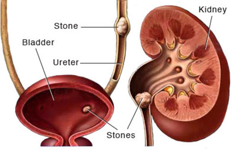
Virtually all stones are formed in the kidneys, initially as small particles. These particles grow within the kidney to varying sizes, often filling up the whole kidney as a branched stone (the staghorn calculus). Sometimes they move out of the kidney when relatively small, and then migrate down the ureter into the bladder. As they migrate down the ureter they may get stuck in any part of ureter and block it causing obstruction to the flow of urine: Larger the stone and higher the location of stone in ureter, higher is the chance of getting it blocked, this results in pain which may be very severe (colic). Ureteric orifices in bladder are very narrow openings around 2-3mm, so stones up to the size of approx 4mm may pass spontaneously. Some reach the bladder, and lodge there, growing larger and larger. Rarely they block the urethra causing a painful retention of urine.

post_989387_1486202152

Symptoms of urinary calculi

Pain : it characteristically starts from the back or lower part of the abdomen and spreads to the entire side . Sometimes it is also referred to the external genitalia and thighs The location and severity of the pain depends upon the location of the stone in the urinary tract, the degree of obstruction, and kidney function. Poorly functioning kidneys cause less pain.


Other symptoms include

  • Blood in urine
  • Fever which indicates infection
  • Urinary trouble
  • Nausea and vomiting
  • Tenderness in the abdomen and on the sides of the spine


Diagnosis

Ultrasound : A widely available investigative modality, detects stones in the kidney with reasonable accuracy. It can also detect the dilated kidney and ureter thus indicating obstruction in the urinary tract. It is less sensitive in localizing ureteral stones, many times fails to see ureteric stones because of abdominal gas.

X-ray KUB : It can detect stones that are radio opaque (i.e. Seen on x-ray). However the accuracy of x-ray depends upon the amount of gases present inside the abdomen and density of the stone.

Intra Venous pyelography(IVP) : This test involves taking a series of x-rays after injecting contrast (dye) into the vein. The contrast flows through the veins and is excreted by the kidneys. As it can be seen on the x-ray, the whole urinary tract containing the contrast can be delineated. Any obstruction in the urinary tract can be picked up. It can also indicate the kidney function and level of stone. A normal kidney function test is a pre requisite to perform IVP.

CT Scan : A non contrast CT scan is the fastest and most accurate investigation for urinary calculi. It can be performed without the need for contrast and thus is safe in patients with history of radio-contrast allergy. It also gives an opportunity to screen other abdominal organs at the same time. Ct scan is now a days investigation of choice for urinary stone disease.


Treatment of kidney stones

Treatment of urinary stones depends upon
  • Size of stone,
  • Number of the stones
  • Location
  • Kidney function
  • Severity of symptoms
  • Presence of infection
  • Anatomy of the urinary tract
Treatment modalities
  • ESWL or Extra-corporeal Shock Wave Lithotrips
  • PCNL or Per Cutaneous Nephrolithotomy
  • URS or Ureterorenoscopy
  • RIRS or Retrograde Intrarenal Surgery
  • Laparoscopic surgery
  • Open surgery


ESWL

ESWL is rarely done now a days.

This procedure focused shockwaves from outside the body to crush stones in the urinary tract. The stone breaks according to its hardness. It is used for stones upto 1 cm in the kidney and upper parts of the ureter. More than one session may be required for breaking the stone. It should not be used in pregnant women. Sometimes the stone pieces get stuck while coming out and may require a ureteroscopic (URS) removal or temporary insertion of a stent. Success rate depends upon size, location and hardness of stone. Overall success rate is only around 60- 70%.

Not a preferred mode of treatment now a days because of highest failure rate and highest stone recurrence rate.

post_667969_1486202717

Mini – PCNL

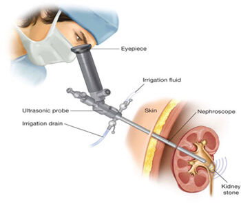
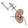
This is one of the most common methods currently employed in the removal of kidney stones. This procedure involves establishing a fine Tract /Puncture / Keyhole from skin into the kidney through which fine Nephroscope (Telescope) is passed inside into kidney and the stone is broken into smaller pieces with Lithoclast/Laser and removed. It can be used for stones of any size. Multiple tracts maybe required for complex/larger stones. The incision used for making these tracts is 5 to 8 mm. and does not require to be stitched usually. Hospitalization is usually required for 2  days and the patient can resume normal work in a weeks time. It has highest success rate approaching upto 95%- 100% for any size of the stone. The only major risk of this operation is bleeding which is unpredictable and can occur in up to 1- 3 % of the patients requiring blood transfusion. Sometimes may require 2nd sitting of PCNL for large stone burden or if unpredictable bleeding occurs.

URS – Uretero-Renoscopic Surgery

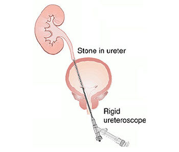
As the name suggests it involves insertion of fine instrument through the penis into the ureter. The stone can be visualized, broken by Lithoclast/ Laser and removed. It is indicated for the treatment of ureteric stones which are stuck up in ureter, obstructing urine flow and causing sever pain, infection or haematuria. Sometimes in the presence of infection or very tight ureter the procedure is done in two stages. A ureteric stent may be required to be placed at the end of the procedure which is removed after two weeks.

URS is a minimal invasive procedure for Ureteric stone with highest success rate. Patient can be discharged same or next day and can resume his duty next.

post_181941_1486202933
RIRS

RIRS – RETRO GRADE INTRA RENAL SURGERY

This is the most advanced and recent technique available for management of small kidney stones.

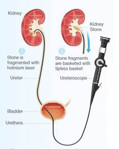
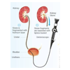
This is a form of uretero-renoscopy, here the instrument used is flexible and thus can negotiate most parts of the ureter and kidney from below. It obviates the need for puncturing the kidney for small  or multiple renal stones.

As it does not requires kidney puncture so there is no risk of bleeding or any risk of kidney injury because of puncture and tracts dilatation. It uses fiber-optics flexible ureteroscope, Laser, laser fibers and delicate instruments, so the  cost of the surgery is higher. It is indicated for smaller (<2cm) kidney and upper Ureteric stones. Success rate depends upon size , number and location of stone. Overall success rate is around 90- 95% but lesser for lower calyceal (pole) stone.

RIRS can be performed in malrotated or ectopic kidney and patients on blood thinners. Even Multiple stones in both the kidneys can also be treated in one sitting. Patients not fit for PCNL surgery can under go RIRS surgery. The only drawback is it might require pre-stenting in few cases before surgery if Ureter is narrow.

Open Surgery

These modalities are rarely used currently, however they have their own place in certain special situations.


Is there a medicine for stones?

There is no known medicine that dissolves urinary stones consistently and predictably. One should drink plenty of fluids, and the doctor will advise you about a specific diet if indicated

post_190372_1486203671

Prevention of stone disease

This is one of the most common methods currently employed in the removal of kidney stones. This procedure involves establishing a tract /puncture from the skin into the kidney through which fine nephroscope (telescope) is passed inside into kidney and the stone is broken into smaller pieces with lithoclast/laser and removed. It can be used for stones of any size. Multiple tracts maybe required for complex/larger stones. The incision used for making these tracts is 5 to 10 mm. and does not require to be stitched usually. Hospitalization is usually required for 2 to 3 days and the patient can resume normal work in a weeks time. It has highest success rate approaching upto 95%- 100% for any size of the stone. The only major risk of this operation is bleeding which is unpredictable and can occur in upto 1- 3 % of the patients requiring blood transfusion. Sometimes may require 2nd sitting of PCNL for large stone burden or if unpredictable bleeding occurs Now a days with advancement of technologies mini-PCNL and micro-PCNL are also done

services_896126_1486031396

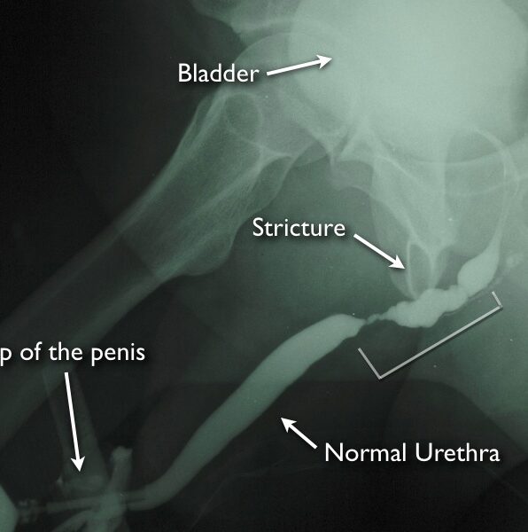
Urethral strictures most commonly result from injury, urethral instrumentation, infection, non-infectious inflammatory conditions of the urethra, and after prior hypospadias surgery. Less common causes include congenital urethral strictures and those resulting from malignancy.

What is the prostate gland?

The prostate is a walnut-sized gland weigh around 20 gram. It is part of male reproductive system. It is located below the urinary bladder and surrounds the upper portion of the urethra (the tube that carries urine out of the body). Its growth is dependent on male hormone Testosterone

What does the prostate do?

The prostate gland is a part of the male reproductive system. It develops at puberty and continues to enlarge throughout life. The prostate gland makes the fluid portion of the semen. This fluid energizes the sperm and makes the vaginal canal less acidic.The seminal vesicles consistent of two pouches that provide nutrients for the sperm and lie immediately behind the prostate. At the point of orgasm , sperm, seminal vesicle fluid and prostatic secretions enter the urethra and mix together, forming semen. This is then ejaculated out through the penis by rhythmic muscular contractions.

What controls the prostate gland?

The growth of the prostate is controlled by testoserone, the male sex hormone. Most testosterone is made by the testicles and a small amount by the adrenal gland. The hormone is converted into dihydro-testosterone (DHT), a more active form that stimulates growth of the gland. The prostate gradually enlarges with ageing, resulting in symptoms such as reduced urine flow and a feeling of incomplete emptying of the bladder having passed urine. This enlargement is usually benign (non-cancerous).

What is prostate cancer?

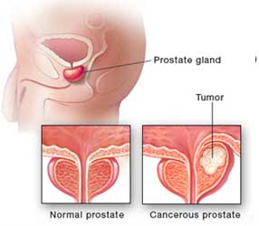
Normally in the prostate, as in the rest of the body, there is a continuous turnover of cells, with new ones replacing old, dying ones. In a cancer, the balance between the new and old cells is lost, with many more new ones being made and older cells living longer, as the process of planned cell death has been disrupted. The malignant growths are known as prostate cancer. They differ from benign enlargements (BPH) in that the cancerous cells can spread (metastasise) to other areas in the body.

post_482973_1485780835

How does prostate cancer spread?

Cancer cell can spread by directly growing outwards through the capsule of the gland into the neighbouring parts of the body, such as the seminal vesicles, bladder or rectum. They may spread through the bloodstream and implant in the bones of the spine and pelvis. Finally, cells can spread through lymphatics to Lymph nodes.

How common is prostate cancer?

Prostate cancer is one of the most common cancer in males, affecting many thousands of men. A man has an estimated 15% chance of developing prostate cancer in his lifetime, that means 1 out of 6 men will develop prostate cancer in their lifetime and a 3% chance of dying from it. Prostate cancer is a disease of men older than 60yrs, rare before 40yrs. Autopsy evidence of prostate cancer is 30% in 4th decade (30s), 50% in 6th decade (50s) and >75% in older than 85yrs.


Why does Prostate Cancer occur

The real answer to this question is not known.Some known risk factors are :
  • African American are the highest risk group of people for prostate cancer.
  • Genetic and familial disease- Relatives of patients with prostate cancer have an increased risk of developing the disease themselves, especially if their father or brother were affected.  Risk doubles if one family member is affected with cancer prostate, if two or more family members are affected risk increases to 7-10 fold.
  • There also appears to be a link with obesity, sedentary life style, high non-vegetarian/ fatty diet and sexually transmitted disease.

What are the symptoms of Prostate Cancer?

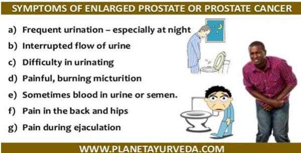
There are often no symptoms associated with early stage prostate cancer and patients may remain unaware of their disease. As the disease progresses and the tumour enlarges, it may press on the urethra and obstruct the flow of urine. Symptoms in early stage are similar to that of (noncancerous) benign enlargement of prostate, like-

  • Weak, interrupted stream of urine
  • Straining
  • On completion he may sill feel that the bladder is not empty.
  • Increased day and night frequency of urination
  • Blood in the semen may be a sign of prostate cancer, although again this is a common finding and not normally related to malignancy.
  • Blood in urine rarely
  • If the tumour is spread to the bones, it may cause pain /backache. Spine is the most common site for this to occur.
  • Decreased appetite and early fatigue-ability.
post_947744_1485781238

How is prostate cancer diagnosed ?

This is the job of a specialist doctor, an Urologist. The doctor will initially ask the patient questions to check their general medical health and see if they are experiencing any symptoms associated with prostate cancer.

post_19344_1485782335

Digital rectal examination

Having made a general examination, the doctor will then need to perform a digital rectal examination to feel the gland. A gloved, lubricated finger is inserted into the rectum to check the size, shape, consistency and any abnormal growth if any of the prostate gland

Blood Test-PSA

The prostate cancer can be evaluated by testing for the level of a particular protein in the blood called PSA (Prostate Specific Antigen). Prostate enlargement tends to cause an increase in the level of PSA, with malignant tumours producing a greater increase than benign enlargements. However, other conditions can also cause PSA to rise, such as a urinary infection or urinary retention. So slight increase in psa level is not specific for prostate cancer.


Ultrasound Examination

The Prostate gland can be imaged with ultrasound for its size, capsule, homogenous/ hetrogenous , any abnormal growth and surrounding  structures like seminal vesicles and bladder


Prostate biopsy

If urologist finds any abnormality in above mentioned examination and tests then he will take the patient for prostate biopsy. Prostate biopsy is done under trasrectal ultrasound guidence or finger guided. With proper preparation under local anaesthesia multiple small pieces of prostate tissue are obtained by a special needle through rectum. The procedure is no more painful than giving an injection, but may occasionally cause a momentary shooting pain in the base of the penis. The doctor will usually give the patient an antibiotic to help prevent any infection occurring.

Between10-12 biopsy cores are normally taken, which are then analysed by histo-pathologist. After the procedure, it is quite common for the patient to see some blood in his urine, semen and stools, but this usually settles over a week or two.


Other Tests

A computer tomography (CT) scan or a magnetic resonance imaging (MRI) scan are sometimes used to obtain pictures of the prostate and the surrounding tissue for staging of prostate cancer.

A bone scan is done to see any bony metastasis.


General Advise/ guidelines

According to American association of urologist (AUA) every men above 55yr should have a urologist consultation, examination and serum PSA test done to rule out prostate cancer and if there is a family history of prostate cancer then at the age of 40 yrs

Stages and Grade of Prostate Cancer

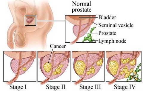
  • The earliest stage, where the cancer is so small that it cannot be felt on rectal examination, but is discovered in a prostate biopsy if raised PSA, or in a prostate tissue that has been surgically removed (as is a transurethral resection of the prostate – TURP) in BPH.
  • The tumour can now be felt on rectal examination, but is still confined to the prostate gland and has not spread.
  • The tumour has spread outside the gland and may have invaded the seminal vesicles.
  • The tumour has spread to involve other surrounding tissues such as rectum, bladder or muscles of the pelvis or bones.
post_45600_1485781645

How is prostate cancer treated?

Treatment of prostate cancer depends upon its stage, grade and patients general condition. Prostate cancer is a very slow growing cancer, many patients live with it without even knowing this. If diagnosed in early stage then after treatment around 90% patients can live normal life for 10 or more years. In western country because of public awareness around 70% patients are diagnosed at early stage but in our country it is just reverse, because of negligence or unawareness more than 70% patients are diagnosed in stage III or IV. There are a number of treatment options for every stage, each with their own advantages and disadvantages. Thus, the therapy needs to be tailored to suit each individual patient. It is possible to cure patients with prostate cancer at an early stage, but in advanced stage when disease is spread in body even if cure is not a possibility, the disease can normally be kept in check for a number of years.


What are the treatment options in prostate cancer?

The different treatment options available for management of prostate cancer are described below. It is important that every patient with such a diagnosis is aware of the different treatments, and they should feel free to discuss these with their Urologist. Whatever therapy is undertaken the patient will need regular follow-up examinations, which may involve a PSA blood test and scans or x-rays, for a number of years.


(A) Active Surveillance and watchful waiting

If the cancer has been discovered accidentally following TURP for BPH or by Biopsy for raised PSA and the patient has no symptoms and disease is localized, “wait and see” policy may be chosen. This does not mean, “do nothing”, but the patient will be regularly monitored by the doctor by digital rectal examination, PSA and may be prostate biopsy and if problems develop or disease progresses appropriate action taken. These actions will often involve the use of hormone therapy, and on such a regime patients commonly live for many years. This choice is most frequently made by those patients with low grade disease specially if elderly.

Advantage : avoid possible side effects of surgery or radiotherapy.

Disadvantage : multiple biopsies and you may miss the chance to   cure the disease

(B) Prostatic Surgery- Radical prostatectomy

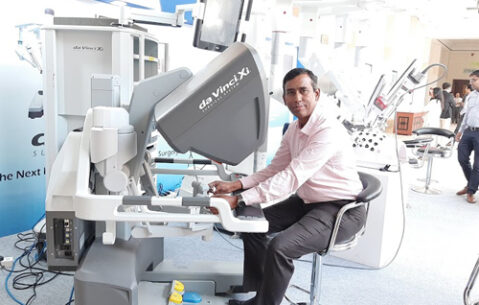
A radical prostatectomy is an operation to remove the entire prostate and seminal vesicles. This operation is performed through an incision in the lower abdomen. This is a complex and major operations that usually require a hospital stay about a week. Such procedures should not be confused with conventional prostate surgery (TURP) done for non cancerous prostate enlargement. The advantage of surgery is that it is one off procedure and provided the cancer is confined to the prostate (stage I&II), will hopefully cure the disease. It avoids side effects of radiotherapy and is considered the most effective form of treatment for early prostatic cancer. However, there are risks associated with radical prostatectomy. It is a major operation, and involves a number of week’s convalescence to make a full recovery. Unfortunately, the prostate lies very close to both the sphincters that control urinary continence and the nerves that produce penile erection. In the past, removal of the gland often caused damage to these structures, resulting in postoperative urinary incontinence and impotence. Newer surgical techniques have reduced the occurrence of impotence and severe incontinence is now less common. Furthermore, there are a number of new therapies to treat such side effects, should they occur. Radical prostatectomy can now be performed with minimal invasive techniques like Laparoscopic RP or Robot assisted RP. Advantage is short convalescence with similar outcome.

ot1
machine

(C) Radiotherapy

Radiotherapy involves high energy radiation aims to destroy the cancerous cells and leave the healthy ones intact. It is a painless procedure, like having an X-ray, although there can be troublesome side effects associated with the treatment. It may be used in two situations: 1. Early cancers confined to the gland or the surrounding tissues 2. For treatment of bony pain in metastatic prostate cancer Radical radiotherapy for a tumour localized to the prostate may be given in two ways. Conventionally, the rays are directed by a machine through the body into the prostate, as with an x-ray. The treatment is given on an out patient basis for five days a week for 4 – 6 weeks. Radiotherapy can also be given using radioactive seeds that are approximately half the size of a grain of rice. These seeds, typically 80-100 in number, are inserted through needles into the prostate. The procedure is performed under anaesthesia. The side effects of radiotherapy are nausea, diarrhea, frequent and painful urination as well as bleeding both in the stools and urine, and local skin reactions. The advantage of radical radiotherapy is that it can cure early prostate cancer without the need of a major surgery. It seldom causes loss of urinary control, and impotence is less common than with surgery.

(D) Hormone Therapy

When the cancer has spread beyond the prostate, going to either the lymph nodes or bones, hormonal therapy is effective at shrinking the tumour, and reducing the side effects of the disease. It does not provide a cure, but may keep the cancer in check for a number of years.

The prostate gland and prostate cancer are under the influence of testosterone, the male sex hormone, which drives the tumour to grow and spread. By blocking the body’s production of testosterone or blocking its action, the growth of the tumour may be greatly reduced. Hormone therapy can be Medical or surgical with similar results.

1)Surgery

The testicles that produce testosterone may be surgically removed by a small operation, called “Orchidectomy” that can be performed as a day care procedure. This has the advantage of being a one off treatment with quickest action, it is most cost effective hormonal treatment.

2)Medical therapy

An injection known as an “LHRH Analogue” is given once a month or every three months and this has a similar effect to removing testicles, but is reversible and does not involve surgery. some medication is prescribed before this to avoid the side effects. Although the result is same but it is a costly treatment and depends on patients compliance.

3)Antiandrogen Tablets

This therapy involves taking daily tablets to block the action of testosterone. The drugs have dual action –

(1) they reduce the production of testosterone by the testicles,

(2) avoid side effects like hot flushes, breast tenderness and impotence. However, Antiandrogen tablets can cause nausea and diarrhea.


(E) Chemotherapy

Chemotherapy is usually in tablet form, involves powerful drugs to attack the cancer cells and try to prevent them growing. It is a second line of defence for patient with advanced stage prostate cancer that is no longer controlled by hormonal therapy

2. URINARY BLADDER CANCER


Urinary bladder cancer

Urinary bladder stores urine formed in kidney, when you urinate, the muscles in the bladder contract and urine is forced out.


What are the layers of the bladder?

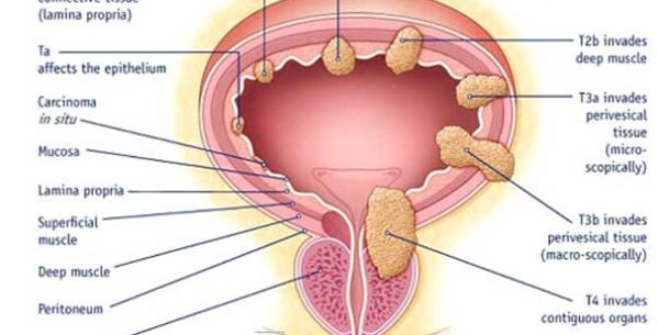
The bladder consists of 3 layers of tissue. The innermost layer which comes into contact with the urine is called the “mucosa” and consists of “transitional cells or urothelium”. These same cells also form the inner lining of the ureters, kidneys and a part of the urethra. The middle layer is known as the “lamina propria”. This layer has a network of blood vessels and nerves and is an important landmark in terms of the staging of bladder cancer. The outer layer of the bladder comprises of the “detrusor muscle” and is called the “muscularis”. Its main function is to relax slowly as the bladder fills up and then to contract to expel the urine out during voiding.

post_643772_1487660663

What is bladder cancer?

Bladder cancer is an uncontrolled abnormal growth and multiplication of cells in the urinary bladder. Most bladder cancers start in the innermost layer of the bladder called the urothelium or transitional epithelium. As the cancer grows into or through the deeper layers in the bladder wall, it becomes more advanced and can be harder to treat. Over time, the cancer might grow outside the bladder and into nearby structures. It might spread to nearby lymph nodes, or to other parts of the body like bones, lungs or liver. Most common type of bladder cancer is Urothelial carcinoma, also known as transitional cell carcinoma (TCC).

Bladder cancers are often described based on how far they have invaded into the wall of the bladder:
  • Non-muscle invasive cancers are still in the inner layer of bladder (mucosa or lamina propria) and have not invaded into the deeper muscle layers.
  • Muscle invasive cancers have grown deeper into muscle layers of the bladder wall. These cancers are more likely to spread and are harder to treat.

What are the causes and risk factors of bladder cancer?

  • Smoking, Tobacco chewing- most common
  • Exposure to toxic chemicals such as arsenic, phenols, aniline dyes, arylamines etc
  • Radiotherapy or chemotherapy for any other disease
  • Long term or chronic infection of bladder
  • Bladder stone or foreign body in bladder


What are bladder cancer symptoms and signs?

  • Bleeding in urine or haematuria- most common.
  • Usually painless and associated with clots.
  • Irritative urinary symptoms like increased frequency, urgency and dysuria


How is bladder cancer diagnosed?

  • Urinanalysis
  • Urine cytology
  • Ultrasonogrphy
  • Cystoscopy and biopsy/ TURBT- This is probably the single most important investigation for bladder cancer. Since there is always a chance to miss small or flat bladder tumors on imaging investigations (ultrasound/CT/MRI) and urine cytology, it is recommended that all patients with bleeding in the urine, without any obvious cause, should have a cystoscopy performed by a urologist as a part of the evaluation and biopsy can be taken.
  • CT Scan

Treatment of bladder tumor

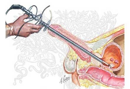
Initial treatment of any bladder tumor is TRANSURETHRAL RESECTION OF BLADDER TUMOR (TURBT), this is done with a special instrument cystoscope introduced through urethra and bladder tumor is resected without any cut or stich on body. This step is most important for confirmation of diagnosis (bladder cancer), staging and grading of tumor.

post_956577_1487661018

Treatment of superficial bladder cancer? (after TURBT)

Small low grade, superficial bladder cancers may not require aggressive management after the initial TURBT and may be simply followed up by doing repeated cystoscopy examinations at regular intervals. It is very important to know that 30-40% of these tumors tend to recur after initial TURBT. High grade, larger, multiple or recurrent superficial bladder cancers may require additional treatment after the initial TURBT. One of the most effective and widely used medications is BCG. It is instilled into bladder through urethra. BCG therapy decreases recurrence and invasiveness of tumor.


Treatment of muscle-invasive bladder cancer?

Surgical removal of the bladder and diversion of the urinary stream using intestinal segments, known as radical cystectomy. This is a major operation.


What is the prognosis for bladder cancer?

The most important factors that impact the prognosis (or the chances of control and cure) of bladder cancer are the Stage and Grade of the tumor. Lower the Stage and Grade, better the outlook

post_630041_1502263981

ERECTILE DYSFUNCTION

Erectile dysfunction (ED) is a condition wherein a man fails to keep an erection firm enough for sexual intercourse. A person with ED have a persistent inability to attain and maintain a sufficient erection so as to permit satisfactory sexual intercourse.Erection trouble at times is not a major cause of concern, however, if the dysfunction is continued for a longer period then it may cause relationship problem, stress or may affect self-confidence.

Epidemiologic studies report that approximately 5–20% of men have moderate to severe erectile dysfunction

Erectile dysfunction may have the following symptoms
  • Continual difficulty in getting an erection
  • Difficulty in maintaining an erection
  • Reduced desire for sexual intercourse

Sexual arousal in men is a complex process that involves the hormones, brain, nerves, emotions, blood vessels and muscles. The brain plays a major part in setting off the series of physical outcomes that makes an erection, which starts with the onset of sexual excitement. Any problem related to the above stated organs or conditions may result in erectile dysfunction.

post_919251_1502264664

Physical causes of erectile dysfunction

A few of the physical causes associated with erectile dysfunction are listed below:
  • High cholesterol
  • Heart disease
  • High blood pressure
  • Clogged blood vessels (atherosclerosis)
  • Low testosterone
Listed below are some of the diseases and conditions that results in EDS:
  • Parkinson’s disease
  • Peyronie’s disease, development of scar tissue inside the penis
  • Multiple sclerosis
  • Diabetes
  • Obesity
  • Certain prescription medications
  • Pelvic area or spinal cord affected by operations or injuries
  • Cancer in prostate cancer and inflamed prostate management
  • Use of Tobacco
  • Substance abuse like alcoholism

Erectile dysfunction (or impotence) is one of the most prevailing complaints in male sexual medicine. Proper diagnosis of the underlying cause of the disease may be enough to treat and reverse erectile dysfunction. ED is becoming quite frequent in diabetes patients. There are mainly two types of ED.Primary, where sexual function is quite normal, or secondary, in which case sexual abnormality occurs after a period of normal sexual activity: in diabetic patients invariably are ED  secondary.

Diabetic men tend to develop ED 10 to 15 years earlier than non- diabetic men. Where approximately 52% of ED is prevalent in the non-diabetic men between the age of 40 to 70 years, in diabetic men it ranges from 35% to 75% that begins at an earlier age. After 70 years of age, 2/3rd of the general male population are diagnosed with ED.

To attain a normal erection, men basically need healthy blood vessels, normal nervous control, proper secretion of male hormones that raises the desire to be sexually stimulated. In diabetic patients blood vessels and nerves that control erection are damaged a person fails to achieve a firm erection despite of normal hormonal secretions and desire to have sex.

Erectile dysfunction in other way is an indication for development of vascular diseases. It is known that the penis acts as a divining rod with its capability to identify individuals at the verge of a vascular catastrophe long before it begins.

Treatments Available for Erectile Dysfunction

Oral drugs: Medications needed for erectile dysfunction include sildenafil, vardenafil or tadalafil. These medicines help ease the flow of blood to the penis, making it easier to attain and maintain firm erection. However, since heart disease is associated with diabetes and ED, THESE DRUGS MAY NOT BE APPROPRIATE IN HEART DISEASE PATIENTS as they cause life threatening interactions with heart medicine. Doctors must be consulted before using these medications.

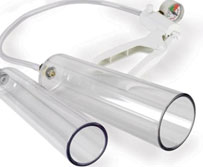
Other treatments for men with ED might consider vacuum constriction devices, intracavernous injection therapy, sex therapy or intraurethral therapy.

post_531843_1502264942

PREMATURE EJACULATION

How does ejaculation occur?

Ejaculation is controlled by the central nervous system and is the combined result of sexual stimulation and impulses resulting from friction that are carried from the spinal cord to the brain.

Ejaculation has two phases:

Phase 1 – Emission

The vas deferens (tubes storing and transporting sperm from the testes), contract to; squeeze the sperm from the prostate gland towards the base of the penis. Seminal vesicles release a secretion that mixes with the sperm to make semen. The ejaculation gets inevitable at this stage.

Phase 2 – Ejaculation

The base muscles of the penis contract and force the semen out of the penis (ejaculation and orgasm) while the bladder neck contracts too. Orgasms may also occur without seminal ejaculation from the penis. A penis will become flaccid after orgasm usually.

What is premature ejaculation?

Premature ejaculation (PE) is caused by the lack of voluntary control over ejaculation. Many men experience ejaculation sooner than they, or their partner, would like during sexual intercourse. PE can often become a frustrating problem and can hinder enjoyment of sex, affect relationships and the quality of life as well. Occasional PE is not a cause for concern however frequent PE can cause distress and strain the couple’s relationship also.

What causes premature ejaculation?

Although the exact cause of premature ejaculation is not known latest research suggests that serotonin, a natural nervous substance, is important. Affecting of serotonin function in the brain may be a cause for PE. These studies have shown that low levels of serotonin in the brain are likely to result in PE.

There are certain psychological factorssuch as temporary depression, stress, abnormal expectations regarding sexual performance, history of sexual repression or low self-confidence can also cause PE. Interpersonal dynamics such as lack of communication between partners, hurt feelings or unresolved disputes also contribute towards achieving sexual intimacy.

Can premature ejaculation occur later in life?

Premature ejaculation (PE) can occur at any age. Although aging is not found to contribute to PE it does result in progressive ejaculation and erectile dysfunction. Erections may be smaller and less hard and they may not be able to hold for a longer time as well.

Can both premature ejaculation and erectile dysfunction affect a man at the same time?

 YES 

When should a doctor be seen?

In case you suffer from premature ejaculation (PE) frequently such that your sexual pleasure gets affected it is advisable to seek a doctor’s advice. To better understand and diagnose the problem the doctor would need to ask you questions, such as:

  • How often does PE occur?
  • How long are you suffering from the problem?
  • Is it limited to one partner? Or is it seen with every partner?
  • Does PE occur at every sexual attempt or just a few?
  • What type of sexual activity (i.e. foreplay, masturbation, intercourse, use of visual clues, etc) is engaged in and how often?
  • How has PE affected sexual activity?
  • What is the quality of personal relationships?
  • How has PE affected your quality of life?
  • Are there any factors that make PE worse or better (i.e. drugs, alcohol, etc)?

How to talk to your partner about premature ejaculation?

Premature ejaculation (PE) affects you, your partner and your sex life as well. The intimacy while intercourse ends abruptly when PE occurs and this might result in anger, shame and frustration in both partners. Communication between partners is considered the best initial step towards diagnosis and treatment.Couple counseling and sex therapy has been found to be affective as well. Different mutual techniques (squeezing technique, etc) may help prolong erection.

What treatments are available?

The treatments for premature ejaculation may include psychological or behavioral therapy and medications. The doctor will be able to advise you the most effective and suitable treatment for you.

Psychological therapies

Psychological therapy can be used singularly or in combination with medical/behavioral therapy for treatment of PE. This therapy helps by recognizing and addressing psychological factors that may be contributing towards PE. This helps the man to decrease inhibitions about sexual performance and increase self-confidence.

Behavioral therapies

Behavioral therapies will suggest effective exercises that require mutual attempts by both partners to prolong ejaculation.
The squeeze method involves the partner to stimulate the man’s penis till near-ejaculation. Just before ejaculation the partner is required to squeeze the penis hard enough to make him lose the erection partially. This exercise aims at helping the man recognize the signs of pre-ejaculation in order to control and delay it on his own.

The start-stop method stimulates the penis till just before ejaculation and abruptly stop the stimulation till the feeling of ejaculation subsides. This process is repeated three times before allowing ejaculation on the fourth time. A repetition of this exercise 3 times/week helps regain control considerably.

Medical therapies

Although not proven much effective anti-depressants and anesthetic creams are often used to treat PE.

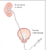
RIRS

RIRS – RETRO GRADE INTRA RENAL SURGERY

This is the most advanced and recent technique available for management of small kidney stones.

This is a form of uretero-renoscopy, here the instrument used is flexible and thus can negotiate most parts of the ureter and kidney from below. It obviates the need for puncturing the kidney for small  or multiple renal stones.

As it does not requires kidney puncture so there is no risk of bleeding or any risk of kidney injury because of puncture and tracts dilatation. It uses fiber-optics flexible ureteroscope, Laser, laser fibers and delicate instruments, so the  cost of the surgery is higher. It is indicated for smaller (<2cm) kidney and upper Ureteric stones. Success rate depends upon size , number and location of stone. Overall success rate is around 90- 95% but lesser for lower calyceal (pole) stone.

RIRS can be performed in malrotated or ectopic kidney and patients on blood thinners. Even Multiple stones in both the kidneys can also be treated in one sitting. Patients not fit for PCNL surgery can under go RIRS surgery. The only drawback is it might require pre-stenting in few cases before surgery if Ureter is narrow.

ThuFLEP – Thulium Fiber Laser Enucleation Of Prostate

 ThuFLEP ( Thulium Fiber Laser Enucleation Of Prostate) is considered world wide new Gold Standard Surgery for enlarged Prostate because of highest success rate. This is most advanced and recent technique for management of enlarged prostate. It has minimum or almost no complications, There is no risk of electrolyte imbalance in it ( which is possible in TURP)

ThuFLEP can be performed in any old age group person, even heart disease persons with pacemaker are not contraindicated. There is negligible or no bleeding and other complications with ThuFLEP. Hospital stay is only 1-2 days. Catheter duration is usually 48 hours.

Laser1
Details of:
  • Laparoscopic Urology Surgeries:-  Lap Pyeloplasty, Lap Nephrectomy, Lap Orchidopexy
  • Laparoscopic & General Surgery: – Lap Cholecystectomy, Lap Hernia, Lap Appendix

+91- 9798900124

Dr Sanjay Jouhary

(MBBS, MS, DNB, Urology)

Details of:

  • Gall Bladder stone
  • Appendix
  • Hernia
  • Hydrocele
  • Piles, Fistula

+91- 9798900124

Dr Sanjay Jouhary

(MBBS, MS, DNB, Urology)

Featured Services

These are our special exams

RIRS

The basis of Wellness

Thu FLEP

Accurate Results

Mini PCNL

With High-End Equipment

Laser URS

Accurate Results

ONE STOP TOTAL UROLOGICAL AND SURGICAL SOLUTION.

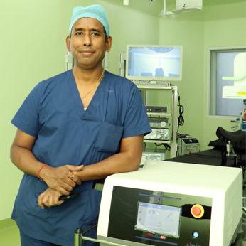
Dr Sanjay Jouhary is one of the leading Urologist, Andrologist, Laser and Laparoscopic Surgeon of Jamshedpur Jharkhand.

Dr Sanjay Jouhary is one of the leading Urologist, Andrologist, Laser and Laparoscopic Surgeon of Jamshedpur Jharkhand. He Has done super-specialization in Urology ( Genitourinary Surgery) from one of the most prestigious institute of eastern India “NH-RABINDRANATH TAGORE HOSPITAL” previously known as “RABINDRANATH TAGORE INTERNATIONAL INSTITUTE OF CARDIAC SCIENCES”- KOLKATA W.B. Dr. Jouhary has been extensively trained in almost all Uro-Surgeries including Kidney transplantation. His areas of special interest are Laser Prostate surgery (ThuFLEP, TURP)  Kidney  & Ureteric stone surgeries ( RIRS, Mini PCNL, Laser URS), Urethroplasty for stricture, Urinary Bladder and Prostate cancer treatment. He has Worked with Lok Nayak Hospital of Maulana Azad Collage New Delhi, Tata Main Hospital Jamshedpur, Tinplate Hospital, Medica Superspeciality Hospital Jamshedpur as Senior Consultant Urologist.  Now he is running his own dedicated “JOUHARY URO AND STONE CLINIC” at 10 Uliyan Main Road Kadma, Jamshedpur since 2008 and  “ JOUHARY URO AND STONE SUPERSPECIALITY HOSPITAL” at 13/1 Kasidih Straight Mile Road, Sakchi  Jamshedpur since 2023.

Integrative Medicine

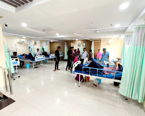
indor1

Want to schedule an appointment?

Call us at +91-9798900124 or fill in the appointment form...